Orthopaedics

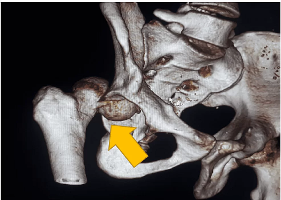
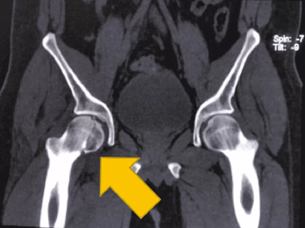
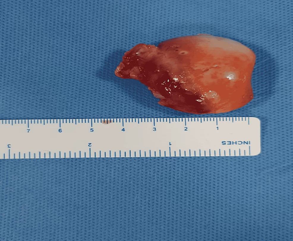
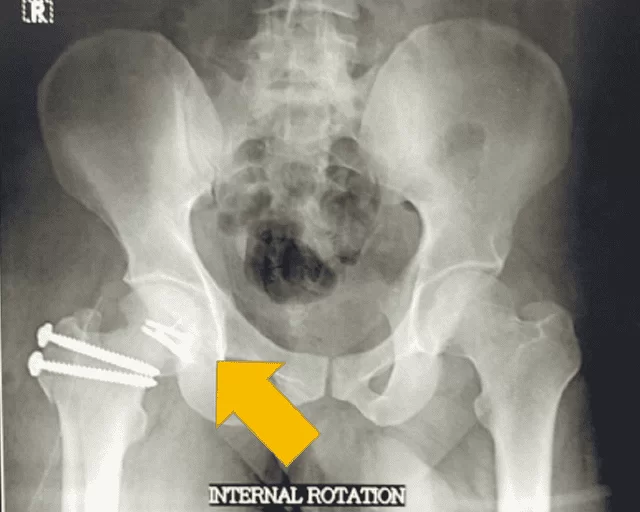
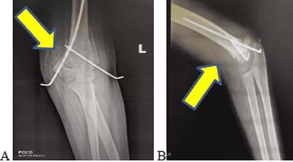
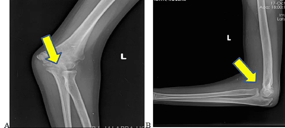
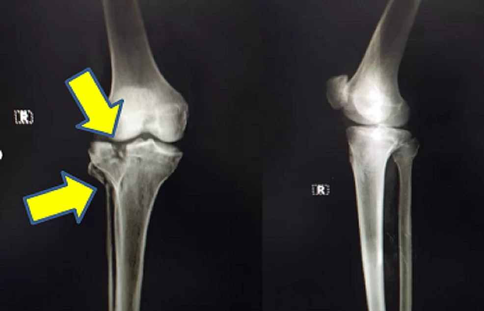
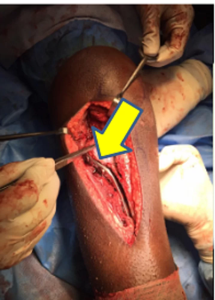
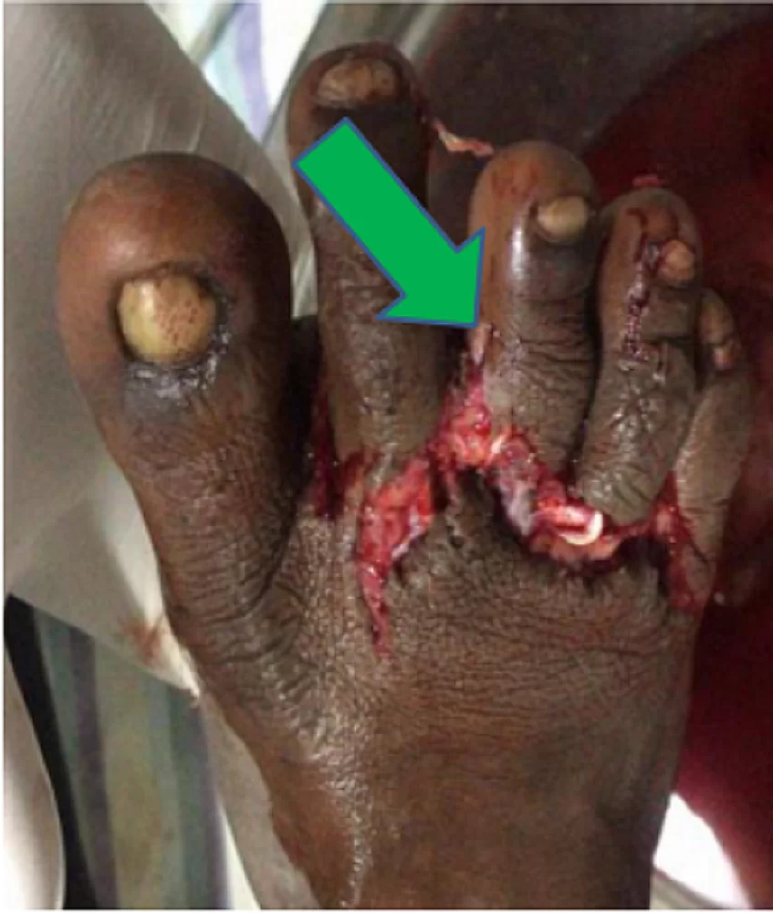
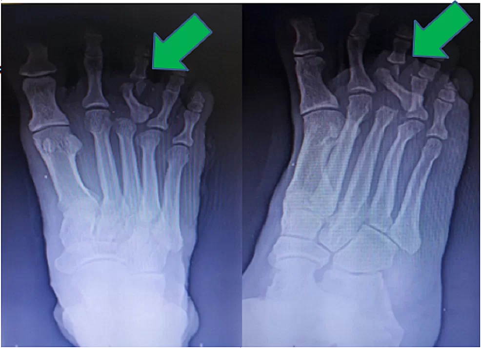
The Department of Orthopaedics was established at R.L. Jalappa Hospital and Research Centre in 1988 and has progressed well from its initial stages, to a fully equipped department with skilled faculty providing compressive trauma care and sub-speciality services. The Dept. has 3 Units and each unit has 2 OPD along with speciality clinics and 2 OT days per week.
Orthopaedic wards have total bed strength of 110. We have exclusive Orthopaedic operation theatres with laminar air flow, with C-Arm’s and all the required instrumentation and implants.

Head of department’s message

It is an honor and privilege to serve as Chair for our storied Department. Our rich history provides a solid foundation for us to further grow our Mission. With reverence to our past, we are looking to our future as leaders of musculoskeletal health care. Earlier this year, the faculty developed a strategic plan to guide us for the next five years. We have quite an ambitious agenda and need support to realize all we would like to do to grow and enhance our mission.
Our department includes fellowship-trained subspecialists in all major areas of orthopedic surgery, including arthroplasty, foot and ankle, spine, pelvis and acetabular surgeries, sports medicine, and orthopedic oncology. Additionally, our team includes highly trained physicians specializing in non-operative treatment of musculoskeletal ailments and injuries. We pride ourselves on a multidisciplinary approach to ensure every one of our patients receives personalized care tailored to their condition and circumstance.
The Department of Orthopedic Surgery is not only committed to providing expert care today but is also dedicated to discovering the most effective musculoskeletal treatments of tomorrow. Every year, eight promising medical students are selected from a pool of over thousands of NEET passed candidates to enter into our three-year comprehensive Orthopaedic surgery training program. Our dedicated faculty are committed to ensuring that future surgeons are prepared to provide the highest level of care to meet their patients' needs in an ever-changing and complex health care system.
When you receive treatment at R L Jalappa hospital and research Centre, your healthcare team will stand by you from diagnosis through post-operative care. Thanks to our residents, physical therapists, social workers, and other specialized doctors, we are poised to lead you on the road to recovery. With a dedication to your long-term well-being, our team will be with you every step of the way as you make your transition from treatment at R L Jalappa hospital and research Centre to your return home and back to your daily life.

Treatments and Procedures

Arthroscopy & Arthroplasty
  • Arthroscopic surgery of various joints, sports medicine and arthroplasty.
  • Arthroscopic ligament reconstruction of ACL & PCL, arthroscopic avulsion fracture fixations
  • Arthroscopic stabilization for recurrent dislocation shoulder, Rotator cuff repair and SLAP tears
  • Arthroscopic meniscal surgeries including meniscal repairs
  • Arthroscopic assisted surgery for recurrent dislocation of patella
  • Complex repairs and reconstructions of multi-ligamentous injuries of knee
  • Joint replacement surgeries of hip, knee and shoulder
  • Primary and revision surgery of hip and knee
  • Management of peri prosthetic fracture, prosthetic joint infection
Complex trauma and spine
  • Polytrauma patient management
  • Management of closed and open fractures
  • Management of complications of fractures and dislocations
  • Acetabular fracture fixations
  • Management of ankle & foot injuries and deformities
  • Minimally invasive procedure for intra-articular fractures
  • Limb reconstruction surgeries
  • Backpain management
  • Stabilization of spine fractures
  • Pedicle screw fixation
Hand and Paediatric surgery
  • Treatment and rehabilitation of traumatised wrist, hand and elbow pathologies.
  • All hand and upper limb fracture management
  • Open carpal tunnel release
  • Trigger finger, De Quervain’s tenosynovitis and tennis elbow surgeries
  • Correction of club foot and DDH
  • Deformity correction of lower limb and limb lengthening
  • Tumour resection and limb reconstruction
  • Deformity correction of knee: High tibial osteotomy
  • Osteomyelitis, septic arthritis and their sequelae management

Facilities and Services

Your browser does not support SVG.
OPD
Minor OT Procedure
Your browser does not support SVG.
IPD
Prstoperative and Surgical intensive care
Your browser does not support SVG.
Emergency Services
Musculoskeletal injuries
Your browser does not support SVG.
Infrastructure
Three Operating Theatres
  • Infrastructure
  • Three operating theatres are equipped with 2 Arthroscopy Instrumentations from Smith & Nephew, Arthrex. There are 4 C-arms of which two are state of the art German made C-arms.

    Operation Theatre is equipped with 8 AO instrumentation sets for carrying out surgeries.

    Saw bone models with instrumentation for training in skill lab.

Faculties

Unit 1
Unit 2
Unit 3
Unit 4

Break through Cases

Fig 1 - Plain radiograph of right hip showed fracture of head of right femur with posterior dislocation of right hip

Fig 2 - Three-dimensional reconstruction CT image showing posterior dislocation with Pipkin’s type II fracture.

Fig 3 - Coronal CT view showing Pipkin’s type II fracture.

Fig 4 - Femur head fracture fragment.

Fig 5 - Intra-operative image of safe surgical dislocation with Pipkin’s fracture reduced with headless cancellous screw fixation.

Fig 6 - Three-dimensional reconstruction CT image showing posterior dislocation with Pipkin’s type II fracture.

Fig 7 - External rotation radiograph of pelvis anteroposterior view showing reduced Pipkin’s fracture at six months follow-up.

Fig 1 - Plain radiograph of left elbow (A) antero-posterior view and (B) lateral view showing left supracondylar fracture with gross rotational anterolateral displacement

Fig 2 - Plain radiographs of the left elbow (A) antero-posterior view and (B) lateral view showing Kirschner wire fixation for left supracondylar fracture with gross rotational anterolateral displacement
Postoperatively, the patient received intravenous amoxicillin-potassium clavulanate twice daily for seven days, followed by oral amoxicillin-potassium clavulanate twice daily for seven days (dosage was based on the weight of the patient). A postoperative radiograph of the operated elbow joint is shown in Figure

Fig 3 - Plain radiographs of the left elbow (A) antero-posterior and (B) lateral views at three months follow-up

Fig 4 - Bilateral carrying angle more in the left side

Fig 5 - Ulnar clawing corrected over the left little finger

Fig 6 - Clinical picture of (A) elbow extension and (B) elbow flexion showing successful functional range of motion after surgical management.

Fig 7 - Plain antero-posterior and lateral left thigh radiograph, indicating non-union of left distal femur with locking compression plate in situ.

Fig 8 - Post-operative left thigh antero-posterior and lateral view radiograph showing removal of the locking compression plate with distal femoral nail fixation with allogenic bone grafting.

Fig 9 - Six-month post-operative, plain antero-posterior left thigh radiograph and lateral view showing fracture uniting with distal femoral nail in place

Fig 1 - Radiographs of Baby 1 & Baby 2

Fig 2 - Strapping Involving Thigh and Leg

Fig 3 - Fracture Unions of Baby 1 & Baby 2

Fig 4 - Lateral ankle radiograph showing bilateral Mulholland deformity

Fig 5 - Postoperative lateral ankle radiograph following surgical excision

Fig 6 - Plain radiographs of the chest with bilateral shoulder anteroposterior view showing bilateral clavicle fracture

Fig 7 - The plain radiograph of the chest with bilateral shoulder anteroposterior view at six months follow up showing locking compression plate in position for left clavicle fracture and united right clavicle fracture

Fig 8 - Clinical picture comparing the range of motion over operative (left) vs. non-operative (right) side

Fig 1 - Plain radiograph (anteroposterior and lateral view) of the right knee showing split fracture involving the lateral tibial condyle, extending into the articular surface, and oblique fracture of the fibular head.

Fig 3 - Plain radiograph (anteroposterior and lateral view) of the right knee showing reduced split fracture involving the lateral tibial condyle with locking compression plate

Fig 1 - Clinical picture of the right foot following trauma.

Fig 2 - Plain radiographs of the right foot in anteroposterior and oblique views showing bipolar dislocation of the proximal phalanx of the third toe and neck of the fourth proximal phalanx fracture.

Fig 3 - Failed closed reduction radiographs.

Fig 4 - Post-reduction following open reduction with Kirschner wire fixation. Hospital course

FAQs
What is orthopaedics?
Orthopaedics is a medical specialty dealing with the diagnosis and treatment of conditions, disorders, and injuries of the muscles, bones, joints, tendons, ligaments, and nerves. A doctor who specializes in this medical specialty is called an orthopaedic surgeon.
What is arthritis?
The word arthritis literally means “joint inflammation.” Arthritis refers to a group of more than 100 rheumatic diseases and other conditions that cause pain, stiffness, and swelling in joints.
Rheumatoid arthritis is a disease that damages the lining surrounding our joints while also destroying our bones, tissue, and joints over time. Osteoarthritis is a progressive condition that slowly damages the cartilage surrounding the ends of bones and is common in the hip, knee, or spine.
What is bursitis?
Bursitis is an inflammation or irritation of a bursa, which is a fluid-filled sac located around joints. Bursitis causes a reduction in or a loss of motion at the affected joint. Bursitis typically occurs in the heel, hip, knee, shoulder, or thumb.
What is cartilage?
Cartilage is a soft, rubbery, gel-like coating on the ends of bones, where they articulate, that protects joints and facilitates movement
What is a ligament?
A ligament is an elastic band of tissue that connects bone to bone and provides stability to the joint
What is a tendon?
A tendon is a band of tissue that connects muscle to bone.
Will I need surgery?
Although orthopedic surgeons are trained and experienced in providing surgery, they do not always prescribe this treatment. Non-surgical options like R.I.C.E. (rest, ice, compression, elevation), casting, physical therapy, and injections can be sufficient in many situations.

Surgical treatment options will vary depending upon your needs and may be used as a last resort.

Arthroscopy, joint replacement, ligament reconstruction, fracture repair, and hand surgery are just some of the most common orthopedic surgeries.
How do injections work for orthopedic health issues?
Orthopedic injections aren’t like numbing shots from the dentist or a flu shot. These can be used for relief from pain, stiffness, and inflammation. Some common conditions they treat include arthritis, nerve pain, and tendonitis. Orthopedic injections can even be used to help diagnose health issues
What is arthroscopic surgery?
This surgery is a non-invasive diagnostic and treatment option that is especially popular in sports medicine. The surgeon will make a small incision with thin needles and use special tools to conduct the procedure. You can have this surgery in a hospital or outpatient facility and it usually doesn’t require more than a day.
What do I need to know about joint replacement surgery?
Joint replacement is a surgical procedure, so it is not for those who could benefit more from non-invasive treatments. During this procedure, the orthopedic surgeon will replace the damaged joint with a prosthesis. These can be ceramic, plastic, or metal.

Doctors typically recommend joint replacement surgery in cases of debilitating joint pain and damage where lifestyle changes, physical therapy, and injections don’t work.

Patient Education material - Click Here