Explore Stories
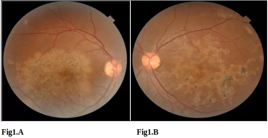
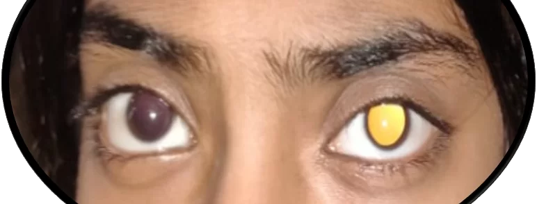
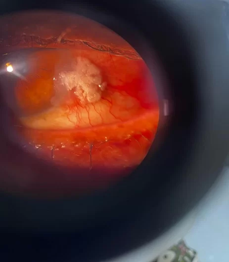
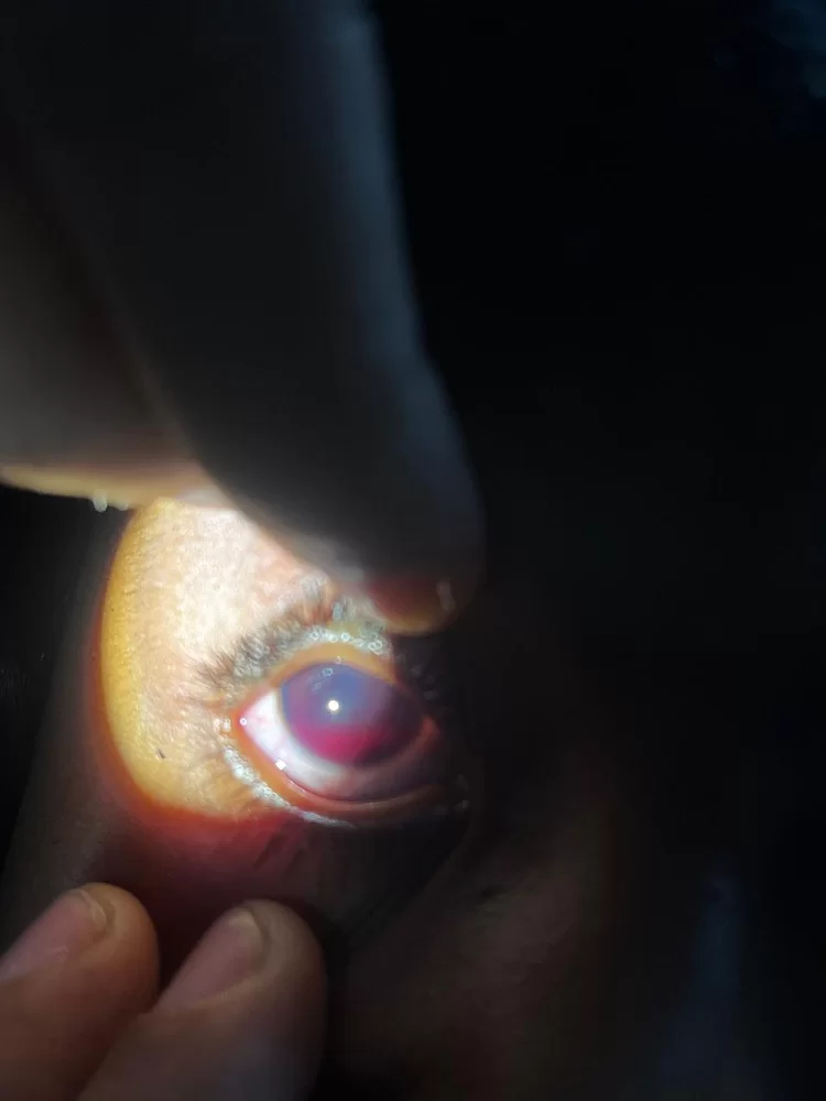
Fig - 1 yellow reflex in LE
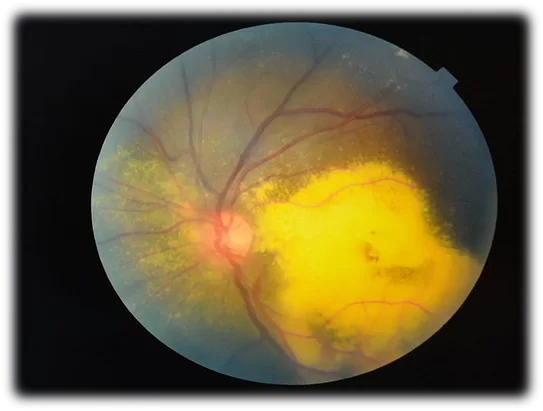
Fig - 2 Extensive subretinal exudates+neovasularization

Fig - 3

Fig - 4
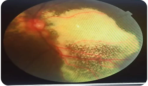
Fig - 5

Fig - 1 showing beaten metal appearance.
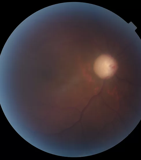
Fig - 2 On Gonioscopy angles are open in BE.
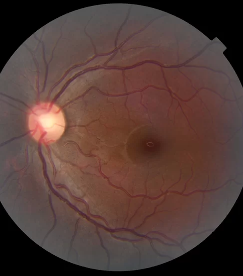
Fig - 3

Fig - OCT RE shows macular thinning
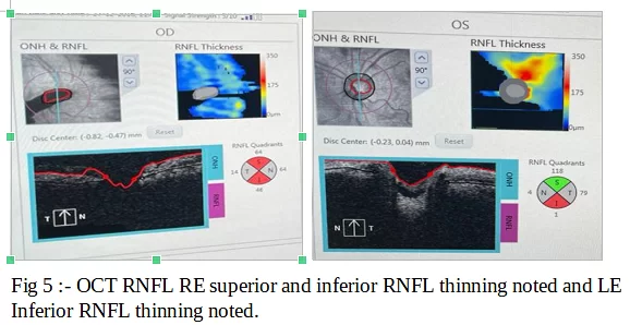
Fig - 5 OCT RNFL RE superior and inferior RNFL thinning noted and LE Inferior RNFL thinning noted.
Preoperative.
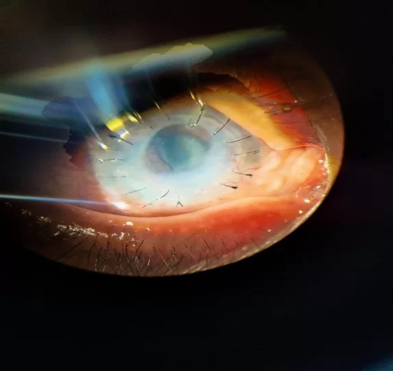
Post operative.
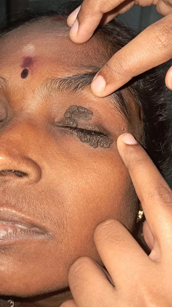
Preoperative.
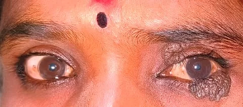
Preoperative
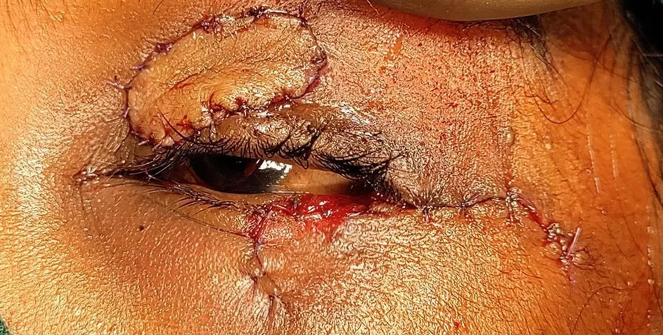
Post operative.
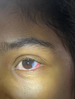
Conjunctival intraepithelial squamous neoplasia.
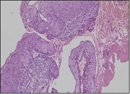
Stratified squamous epithelium with full thickness dysplasia
Excision with cryotherapy with Mitomycin C
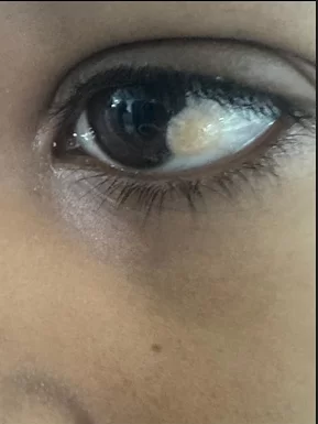
Preoperative.
Post operative
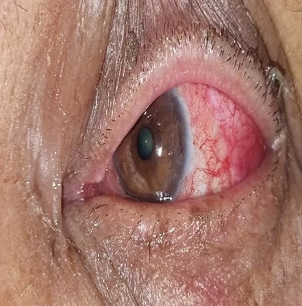
Clinical OSSN seen as well-demarcated corneal epithelial clouding emanating from the head of the pterygium.
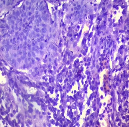
Focal areas show moderate dysplasia suggestive of Conjunctival Squamous Intraepithelial neoplasia
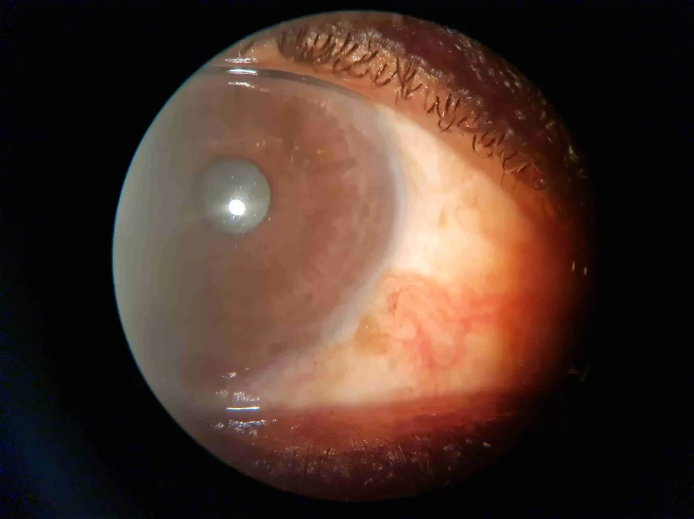
Excision with cryotherapy with Mitomycin C Postoperative follow up at 8 months
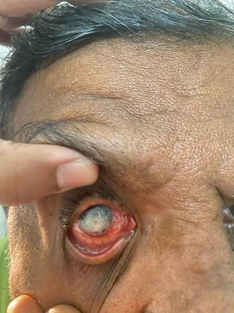
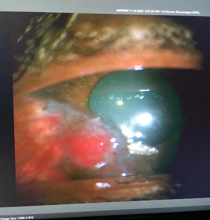
Hyphema .
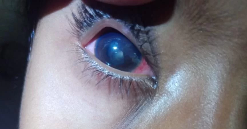
Complicated cataract
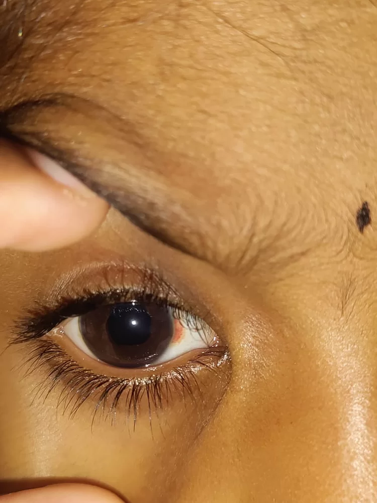
Post-surgery with 6/6 p VA