Break through Cases
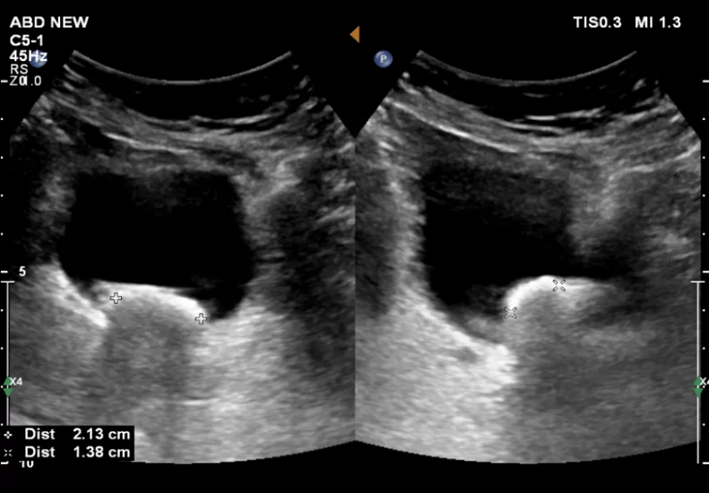
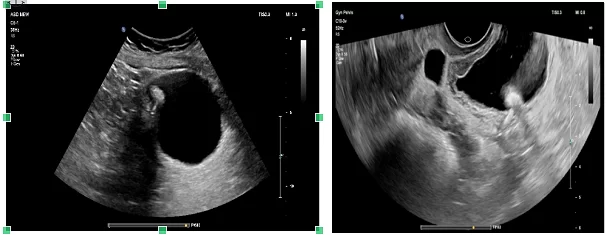
Two calculi measuring ~ 2.1 cm & 1.4 cm in dependent part of bladder. Wall appears thickened with mucosal irregularities and internal echogenic debris.
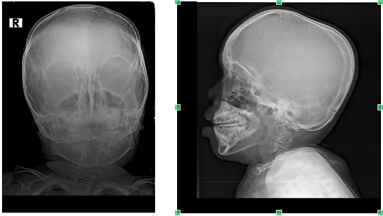
RADIOGRAPH OF SKULL AP & LATERAL VIEW
Shows mild calvarial thickening with normal facial bones.
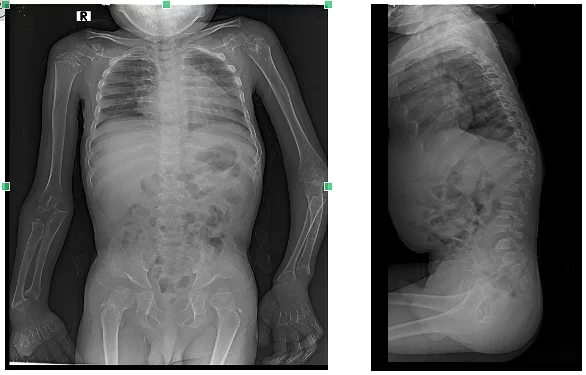
RADIOGRAPH WHOLE SPINE AP AND LATERAL VIEWS
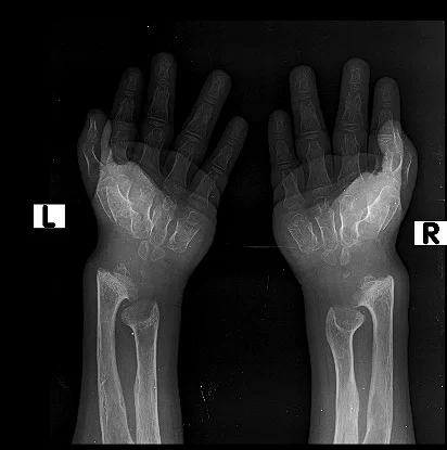
RADIOGRAPH BILATERAL HANDS WITH WRIST AP VIEW
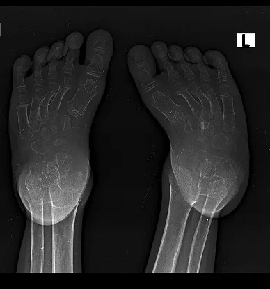
RADIOGRAPH BILATERAL FOOT AP VIEW
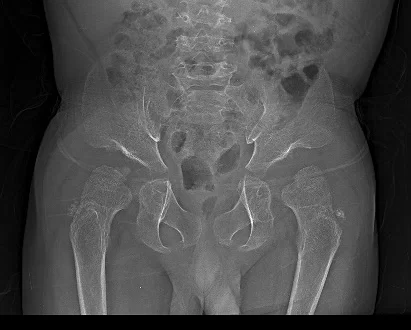
Widening of neck of femur